Comprehensive Histopathological Study of Postmortem Renal Tissues: A Retrospective Study in Baghdad (2020-2024)
الكلمات المفتاحية:
Autopsy، Kidney، Pyelonephritis، Postmortem، Renal pathologyالملخص
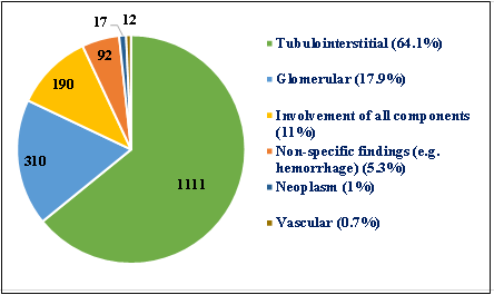
Background: Autopsy represents a comprehensive method for direct macroscopic and microscopic assessment of organs following death. Despite progress in imaging and molecular studies, postmortem renal histopathological examination continues to be the most reliable method for precise identification of renal diseases. Objective: To determine the frequency of renal diseases in autopsy cases, identify pathological findings, and assess their association with demographic factors. Methods: A retrospective study was conducted over one year on 1,834 postmortem renal tissue samples obtained from autopsies performed between 2020 and 2024 at the Directorate of Forensic Medicine, Baghdad, Iraq. Renal lesions were classified as tumor, glomerular, tubular, interstitial, and vascular. Results: Autolysis was present in 102 cases. Out of the 1,732 autopsy cases, 73% were male, with an average age of 52.8±18.4 years. Hemorrhagic foci were the most prevalent gross finding (72.9%), with tubulointerstitial lesions being the most common histological pattern (64.1%). Pyelonephritis was the most common microscopic diagnosis (40.8%). Renal cell carcinoma was the most prevalent neoplasm (52.9%). Renal diagnoses were significantly associated with age and sex (p<0.001). Conclusions: Males were more likely to develop renal pathological abnormalities, which increased as they aged. The prevalence of hemorrhagic findings indicates a high incidence of acute and inflammatory kidney diseases, with pyelonephritis being the most common microscopic diagnosis. The discovery of malignant tumors demonstrated the value of postmortem renal histopathology investigation in identifying previously unknown illnesses.
التنزيلات
المراجع
1. Sulegaon R, Kulkarni D, Chulki S. Medicolegal autopsies-Interesting and incidental findings. Int J Forensic Sci Pathol. 2015;3(8):156-160. doi: 10.19070/2332-287X-1500038.
2. Ramos SG, Ottaviani G, Peres LC, Rattis BAC, Leão PS, Akel TN, et al. Why should clinical autopsies continue to exist? Diagnostics (Basel). 2021;11(8):1482. doi: 10.3390/diagnostics11081482.
3. Xie K, Cao H, Ling S, Zhong J, Chen H, Chen P, et al. Global, regional, and national burden of chronic kidney disease, 1990-2021: a systematic analysis for the Global Burden of Disease Study 2021. Front Endocrinol (Lausanne). 2025;16:1526482. doi: 10.3389/fendo.2025.1526482.
4. Batte A, Shahrin L, Claure-Del Granado R, Luyckx VA, Conroy AL. Infections and acute kidney injury: A global perspective. Semin Nephrol. 2023;43(5):151466. doi: 10.1016/j.semnephrol.2023.151466.
5. Couser WG, Remuzzi G, Mendis Su, Tonelli M. The contribution of chronic kidney disease to the global burden of major noncommunicable diseases. Kidney Int. 2011;80(12):1258-1270. doi: 10.1038/ki.2011.368.
6. Ali AS, Al-Saedi AJ. Renal biopsy practice in Iraq: a systematic review. Iraqi J Med Sci. 2016;14(4):393-399. doi: 10.22578/IJMS.14.4.13.
7. Obrycki Ł, Sarnecki J, Lichosik M, Sopińska M, Placzyńska M, Stańczyk M, et al. Kidney length normative values in children aged 0-19 years - a multicenter study. Pediatr Nephrol. 2022;37(5):1075-1085. doi: 10.1007/s00467-021-05303-5.
8. Garza FA, Leslie SW. Anatomy, Abdomen and Pelvis: Kidneys. In: StatPearls [Internet]. Treasure Island (FL): StatPearls Publishing; 2025 [cited 2026 Oct 8]. Available from: https://www.ncbi.nlm.nih.gov/books/NBK482385
9. Castelino K, Devaraju S, Bhat A, Thattil A. The morphological differences between acute tubular necrosis and autolysis: An autopsy study. J Indian Acad Forensic Medi. 2023;45(4):400-403. doi: 10.48165/jiafm.2023.45.4.21.
10. Desai K, Mehta N, Goswami H. Histomorphological spectrum of kidney lesions in autopsy cases. Int J Contemp Pathol. 2020;6(1):29-33. doi: 10.37506/ijcpath.v6i1.11914.
11. Omenai SA, Ajani MA, Nwadiokwu JI, Okolo CA. Histomorphological assessment of non-neoplastic renal diseases at autopsy: an institutional experience in Southwestern Nigeria. Malawi Med J. 2021;33(4):281-286. doi: 10.4314/mmj.v33i4.9.
12. Perrone ME, Chang A, Henriksen KJ. Medical renal diseases are frequent but often unrecognized in adult autopsies. Mod Pathol. 2018;31(2):365-373. doi: 10.1038/modpathol.2017.122.
13. Basra S, Singh JP. Autopsy analysis of renal lesions: a histomorphological perspective. J Forensic Med Toxicol. 2025;42(2):40-47. doi: 10.48165/jfmt.2025.42.2.8.
14. Padmanabhan A, Gohil S, Gadgil NM, Sachdeva P. Chronic renal failure: An autopsy study. Saudi J Kidney Dis Transpl. 2017;28(3):545-551. doi: 10.4103/1319-2442.206441.
15. Khare P, Gupta R, Agarwal S, Bhatnagar A, Anand R. Spectrum of renal lesions on autopsy: Experience of a tertiary level institute based on retrospective histopathological analysis. Cureus. 2021;13(8):e17064. doi: 10.7759/cureus.17064.
16. Ayukekbong JA, Ntemgwa M, Atabe AN. The threat of antimicrobial resistance in developing countries: causes and control strategies. Antimicrob Resist Infect Control. 2017;6:47. doi: 10.1186/s13756-017-0208-x.
17. Sandhu VK, Puri A, Singh N. The histomorphological spectrum of renal lesions in an autopsy study. Ann Pathol Lab Med. 2017;4(4):A410-414. doi: 10.21276/APALM.1455.
18. Saniha PB, Parvathaneni S, Sneha K, Jadav MN. Morphological spectrum of kidney lesions-an autopsy study. J Chem Health Risks. 2025;15(2):767-772. Available at: https://jchr.org/index.php/JCHR/article/view/7886?utm_source
19. Sahoo SK, Mishra J, Perei S, Sahu A. Histopathological spectrum of kidney lesions in autopsy: a teaching hospital based study. Int J Acad Med Pharm. 2023;5(5):945-951. doi: 10.47009/jamp.2023.5.5.185.
20. Mondal S, Pradhan R, Chatterjee S, Biswas S, Sikder M. Evaluation of chronic kidney disease in sudden death cases: a one year autopsy study in tertiary care hospital. J Indian Acad Forensic Med. 2023;45(1):41-44. doi: 10.48165/jiafm.2023.45.1.11.
21. Pais P, Iyengar A. Kidney care for all: Addressing the gaps in an imperfect world-A global perspective on improving access to kidney care in low-resource settings. Kidney360. 2023;4(7):982-986. doi: 10.34067/KID.0000000000000128.
22. Klessens CQ, Woutman TD, Veraar KA, Zandbergen M, Valk EJ, Rotmans JI, et al. An autopsy study suggests that diabetic nephropathy is underdiagnosed. Kidney Int. 2016;90(1):149-156. doi: 10.1016/j.kint.2016.01.023.
23. Abusaib M, Ahmed M, Nwayyir HA, Alidrisi HA, Al-Abbood M, Al-Bayati A, et al. Iraqi experts consensus on the management of type 2 diabetes/prediabetes in adults. Clin Med Insights Endocrinol Diabetes. 2020;13:1179551420942232. doi: 10.1177/1179551420942232.
24. Rzepecka-Woźniak E, Trzepla A, Okoń K. Renal tumors in unselected forensic post-mortem material. Pol J Pathol. 2014;65(3):218-22. doi: 10.5114/pjp.2014.45784.
25. Dharshini GV, Jeyalakshmi Devi N. Histopathological spectrum of kidney lesions in autopsies from a tertiary care hospital. Int J Med Sci Curr Res. 2024;7(5):539-543. Available at: https://ijmscr.com/isset/images/uploads/17302691327676.pdf?utm_sourc
26. Mulayamkuzhiyil Saju J, Leslie SW. Renal Infarction. 2024 Mar 10. In: StatPearls [Internet]. Treasure Island (FL): StatPearls Publishing; 2024 Jan–. PMID: 35881744.
27. Sumnu A, Gursu M, Ozturk S. Primary glomerular diseases in the elderly. World J Nephrol. 2015;4(2):263-270. doi: 10.5527/wjn.v4.i2.263.
28. Kattah AG, Garovic VD. Understanding sex differences in progression and prognosis of chronic kidney disease. Ann Transl Med. 2020;8(14):897. doi: 10.21037/atm.2020.03.62.
29. Oshima M, Iwata Y, Toyama T, Kitajima S, Hara A, Sakai N, et al. Sex differences in risk factors for end-stage kidney disease and death in type 2 diabetes: a retrospective cohort study. J Diabetes. 2023;15(3):246-254. doi: 10.1111/1753-0407.13367.
التنزيلات
منشور
كيفية الاقتباس
إصدار
القسم
الرخصة
الحقوق الفكرية (c) 2026 Al-Rafidain Journal of Medical Sciences

هذا العمل مرخص بموجب Creative Commons Attribution-NonCommercial-ShareAlike 4.0 International License.
Published by Al-Rafidain University College. This is an open access journal issued under the CC BY-NC-SA 4.0 license (https://creativecommons.org/licenses/by-nc-sa/4.0/).