Mechanistic Divergence in Chemotherapy-Induced Genotoxicity: A Comparative in vivo Assessment of Different Antineoplastic Drug Classes in Murine Bone Marrow and Spleen
الكلمات المفتاحية:
Antineoplastic agents، Chromosome aberrations، Genotoxicity، Micronucleus tests، Mitotic indexالملخص
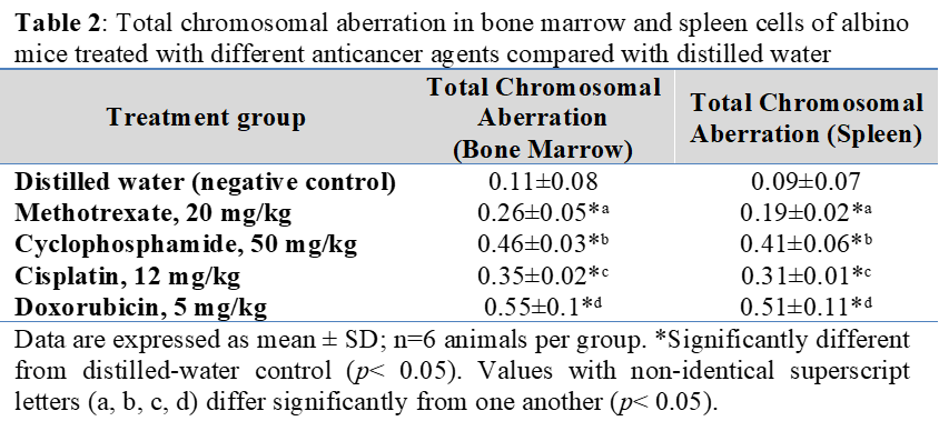
Background: Cancer chemotherapeutic agents, while effective against malignant cells, can also damage rapidly dividing normal tissues such as the bone marrow, raising concern about their genotoxic potential. Objective: To compare the acute in vivo cytotoxic and genotoxic effects of four mechanistically distinct antineoplastic agents on the bone marrow and splenic cells of albino mice. Methods: Sixty albino mice were randomly allocated to five groups (n=12): a negative control receiving distilled water and four groups receiving a single intraperitoneal dose of cisplatin (12 mg/kg), methotrexate (20 mg/kg), doxorubicin (5 mg/kg), or cyclophosphamide (50 mg/kg). Twenty-four hours after treatment, bone marrow and spleen samples were evaluated for mitotic index (MI), total chromosomal aberrations (TCA), and micronucleus (MN) frequency. Results: All four agents significantly reduced MI and increased TCA and MN frequency in both tissues compared with the control (p<0.05). Methotrexate produced the smallest changes across all endpoints, whereas cyclophosphamide and cisplatin produced moderate, distinct effects. Doxorubicin caused the greatest reduction in MI and the highest TCA and MN frequencies. Conclusions: Genotoxicity ranked doxorubicin > cyclophosphamide > cisplatin > methotrexate, inversely reflecting the antiproliferative potency of these agents and paralleling the hematological toxicity observed clinically.
التنزيلات
المراجع
Chen J, Potlapalli R, Quan H, Chen L, Xie Y, Pouriyeh S, et al. Exploring DNA damage and repair mechanisms: a review with computational insights. BioTech (Basel). 2024;13(1):3. doi: 10.3390/biotech13010003.
Roos WP, Kaina B. DNA damage-induced cell death: from mechanisms to clinical applications. Cancer Lett. 2024;582:216524. doi: 10.1016/j.canlet.2023.216524.
Jackson SP, Bartek J. The DNA-damage response in human biology and disease. Nature. 2024;625(7994):245-256. doi: 10.1038/s41586-023-06842-0.
Smith JA, Redon CE, Aladjem MI. Cellular senescence and genomic instability induced by chemotherapy: a comprehensive review. Cell Death Dis. 2025;16(2):112. doi: 10.1038/s41419-025-07412-1.
Martinez-Pastor B, Mostoslavsky R. Chromatin dynamics in response to DNA damage and its impact on tissue homeostasis. Trends Cell Biol. 2025;35(3):201-215. doi: 10.1016/j.tcb.2024.10.004.
Gartner A, Engebrecht J. DNA repair, recombination, and damage signaling. Genetics. 2022;220(4):iyab178. doi: 10.1093/genetics/iyab178.
Sasanuma H, Takeda S. Mechanisms maintaining genomic stability: pathways repairing DNA double-strand breaks. Int J Mol Sci. 2024;25(4):2105. doi: 10.3390/ijms25042105.
OECD. Test No. 487: In Vitro Mammalian Cell Micronucleus Test. OECD Guidelines for the Testing of Chemicals, Section 4. Paris: OECD Publishing; 2023. doi: 10.1787/9789264264861-en.
Di Leonardo A, Fardilha M, Gorbsky GJ. Chromosomal instability and micronuclei formation in cancer progression. Mutat Res. 2025;831:111952. doi: 10.1016/j.mrfmmm.2025.111952
Santaguida S, Amon A. Short- and long-term consequences of chromosome mis-segregation and micronuclei. Nat Rev Mol Cell Biol. 2024;25(1):19-36. doi: 10.1038/s41580-023-00659-1.
Molehin OR, Afolabi AT. Genotoxicity of cancer therapies and the risk of secondary malignancies: toward personalized prevention. Clin Transl Oncol. 2026;28(1):45-56. doi: 10.1007/s12094-026-04227-6.
Kennedy RE, Lord CJ. Mechanisms of genotoxicity induced by conventional chemotherapy and targeted agents. Lancet Oncol. 2025;26(4):e155-e167. doi: 10.1016/S1470-2045(25)00021-X.
Chan BS, Bosco AA, Buckley NA. Navigating methotrexate toxicity: examining the therapeutic roles of folinic acid and glucarpidase. Br J Clin Pharmacol. 2025;91(3):628-635. doi: 10.1111/bcp.16096.
Visentin M, Zhao R, Goldman ID. The antifolates: essential mechanisms of transport and action. Mol Pharmacol. 2024;105(2):89-101. doi: 10.1124/molpharm.123.000781.
Smyrniotou A, Castana O, Kartalou M. Methotrexate-induced oxidative stress and cellular senescence in human vascular endothelium. Toxicology. 2024;499:153671. doi: 10.1016/j.tox.2024.153671.
Mahmoud AM, Hozayen WG. Oxidative mechanisms underlying methotrexate genotoxicity: protective role of natural antioxidants. Environ Sci Pollut Res. 2025;32(8):10412-10427. doi: 10.1007/s11356-025-36109-w.
Dabbish E, Scoditti S, Shehata MNI, Ritacco I, Ibrahim MAA, Shoeib T, et al. Insights on cyclophosphamide metabolism and anticancer mechanism of action: a computational study. J Comput Chem. 2024;45(10):663-670. doi: 10.1002/jcc.27280.
Emadi A, Jones RJ, Brodsky RA. Cyclophosphamide and its mechanisms of action in conventional and high-dose therapy. Blood Rev. 2024;63:101123. doi: 10.1016/j.blre.2023.101123.
Yadav V, Krishnan A, Zahiruddin S, Ahmad S, Vohora D. Amelioration of cyclophosphamide-induced DNA damage, oxidative stress, and hepato- and neurotoxicity by Piper longum extract in rats: the role of γH2AX and 8-OHdG. Front Pharmacol. 2023;14:1147823. doi: 10.3389/fphar.2023.1147823.
Abdel-Latif S, El-Shazly S. Cytogenetic evaluation of cyclophosphamide-induced damage in rodent bone marrow models: an updated protocol. Mutat Res Genet Toxicol Environ Mutagen. 2025;894:503712. doi:10.1016/j.mrgentox.2025.503712
Gandin V, Hoeschele JD, Margiotta N. Cisplatin in cancer therapy: molecular mechanisms of action 3.0. Int J Mol Sci. 2023;24(9):7917. doi: 10.3390/ijms24097917.
Rottenberg S, Bluzer N. Platinum resistance in cancer: molecular mechanisms and cellular processing of cisplatin-DNA adducts. Drug Resist Updat. 2024;73:101042. doi: 10.1016/j.drup.2024.101042.
Qiu Y, Lin X, Mladenov E, Mladenova V, Thomale J, Sak A, et al. Cell-cycle-specific lesion evolution rather than inhibition of double-strand-break repair underpins cisplatin radiosensitization. Mol Oncol. 2026;20(7):1452-1470. doi: 10.1002/1878-0261.70240.
Bhutani V, Varzideh F, Wilson S, Kansakar U, Jankauskas SS, Santulli G. Doxorubicin-induced cardiotoxicity: a comprehensive update. J Cardiovasc Dev Dis. 2025;12(6):207. doi: 10.3390/jcdd12060207.
Dubey N, Singh S, Panchbhai P, Chaturvedi G, Goyal A, Singh N, et al. Doxorubicin-induced cardiotoxicity: molecular mechanisms to novel therapeutic frontiers of recent preclinical discoveries. Cell Biochem Biophys. 2026;84(2):345-362.
Khynriam D, Prasad SB. Cisplatin-induced genotoxic effects and endogenous glutathione levels in mice bearing ascites Dalton's lymphoma. Mutat Res. 2003;526(1-2):9-18. doi: 10.1016/S0027-5107(03)00005-8.
Hassan AF, Numan IT, Hamad MN. Evaluation of genotoxicity and effect of petroleum ether extract of Euphorbia lathyris on bone marrow cells: in-vivo study. Int J Pharm Sci Rev Res. 2015;32(2):116-120. doi: 10.3923/ijcr.2018.86.91.
Hajra S, Patra AR, Basu A, Bhattacharya S. Prevention of doxorubicin (DOX)-induced genotoxicity and cardiotoxicity: Effect of plant derived small molecule indole-3-carbinol (I3C) on oxidative stress and inflammation. Biomed Pharmacother. 2018;101:228-243. doi: 10.1016/j.biopha.2018.02.088.
Kour J, Ali MN, Ganaie HA, Tabassum N. Amelioration of the cyclophosphamide induced genotoxic damage in mice by the ethanolic extract of Equisetum arvense. Toxicol Rep. 2017;4:226-233. doi: 10.1016/j.toxrep.2017.05.001.
Radhi A, Al-Shawi N, Hassan A. Impact of omega-3 alone or in combination with irinotecan on bone marrow and spleen of rats: in vivo study. Iraqi J Pharm Sci. 2023;32(1):53-58. doi: 10.31351/vol32iss1pp53-58.
Hayashi M. The micronucleus test-most widely used in vivo genotoxicity test. Genes Environ. 2016;38:18. doi: 10.1186/s41021-016-0044-x.
Tice RR, Hayashi M, MacGregor JT, Anderson D, Blakey DH, Holden HE, et al. Report from the working group on the in vivo mammalian bone marrow chromosomal aberration test. Mutat Res. 1994;312(3):305-312. doi: 10.1016/0165-1161(94)00016-6.
التنزيلات
منشور
كيفية الاقتباس
إصدار
القسم
الرخصة
الحقوق الفكرية (c) 2026 Al-Rafidain Journal of Medical Sciences

هذا العمل مرخص بموجب Creative Commons Attribution-NonCommercial-ShareAlike 4.0 International License.
Published by Al-Rafidain University College. This is an open access journal issued under the CC BY-NC-SA 4.0 license (https://creativecommons.org/licenses/by-nc-sa/4.0/).