Belatacept Normalizes Specific Epigenetic-Cytokine Crosstalk in an Animal Model of Concanavalin A-Induced Acute Hepatic Injury
DOI:
https://doi.org/10.54133/ajms.v11i1.3026الكلمات المفتاحية:
Belatacept، Interleukin-1β ، Liver injury ، MiRNA-155 ، MiRNA-223الملخص
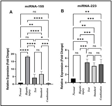
Background: Acute liver injury is still a major therapeutic problem. Studies have focused on the epigenetic environment that controls liver injury. MicroRNAs have become essential post-transcriptional regulators for the production of pro-inflammatory signaling cascades. Objective: This study evaluated the therapeutic potential of belatacept in hepatic injury by measurement of hepatic miRNA-155 and miRNA-223 and assessed its impact on IL-1β expression. Methods: Concanavalin-A was used to induce liver injury in Wistar rats, which were treated with prednisolone, belatacept, or a combination therapy. Hepatic expression of targeted miRNAs was quantified using RT-qPCR. Hepatic IL-1β was measured by the Western blotting technique. Results: The findings of this study identified that belatacept significantly normalized the expression of miRNA-155 and miRNA-223 (p<0.05). This epigenetic recovery was correlated with the stabilization of hepatic IL-1β level (p<0.01), Furthermore, belatacept demonstrated a therapeutic potency superior to prednisolone in mitigating liver injury, while combination therapy showed no significant synergistic advantage, suggesting a saturation of the common protective pathways. Conclusions: Belatacept ameliorates hepatic injury by targeting the miRNA-155/Akt/IL-1β pathway. Overall, miRNA-155 may offer a successful biomarker for treatment monitoring, and the miR-155/Akt pathway is a promising target for improving management of liver injury.
التنزيلات
المراجع
Fadhil AF, Kamal YM, Waheed HJ. The hepatoprotective effect of Glycyrrhiza glabra roots extract against amiodarone-induced hepatotoxicity in male rats. Iraqi J Pharm Sci. 2024;33(2):190–196. doi: 10.31351/vol33iss2pp190-196 DOI: https://doi.org/10.31351/vol33iss2pp190-196
Hameed FG, Arif IS, Mohammed MM, Arif HS. Co-occurrence of Wilson disease and autoimmune hepatitis in a 14-year-old female: a case report. Al Mustansiriyah J Pharm Sci. 2021;21(2):1–8. doi: 10.32947/ajps.v21i2.803 DOI: https://doi.org/10.32947/ajps.v21i2.803
Diener C, Keller A, Meese E. The miRNA–target interactions: an underestimated intricacy. Nucleic Acids Res. 2024;52(4):1544–1557. doi: 10.1093/nar/gkad1142. DOI: https://doi.org/10.1093/nar/gkad1142
Almajidi YQ, Muslim RK. Oligonucleotides as a novel therapeutic approach: an innovative area for drug delivery in neurological disorders. Al Mustansiriyah J Pharm Sci. 2025;25(1):143–169. doi: 10.32947/ajps.v25i1.1065. DOI: https://doi.org/10.32947/ajps.v25i1.1065
Gehrke N, Hövelmeyer N, Waisman A, Straub BK, Weinmann-Menke J, Wörns MA, et al. Hepatocyte-specific deletion of IL1-RI attenuates liver injury by blocking IL-1-driven autoinflammation. J Hepatol. 2018;68(5):986–995. doi: 10.1016/j.jhep.2018.01.008. DOI: https://doi.org/10.1016/j.jhep.2018.01.008
Diep S, Maddukuri M, Yamauchi S, Geshow G, Delk NA. Interleukin-1 and nuclear factor kappa B signaling promote breast cancer progression and treatment resistance. Cells. 2022;11(10):1673. doi: 10.3390/cells11101673. DOI: https://doi.org/10.3390/cells11101673
Craig R, McIntosh K, Ho KH, McCulloch A, Riley C, Lawson C, et al. IL-1β stimulates a novel axis within the NF-κB pathway in endothelial cells regulated by IKKα and TAK-1. Biochem Pharmacol. 2025;232:116736. doi: 10.1016/j.bcp.2024.116736. DOI: https://doi.org/10.1016/j.bcp.2024.116736
Mao H, Zhao X, Sun SC. NF-κB in inflammation and cancer. Cell Mol Immunol. 2025;22(8):811–839. doi: 10.1038/s41423-025-01310-w. DOI: https://doi.org/10.1038/s41423-025-01310-w
Cao YY, Wang Z, Wang ZH, Jiang XG, Lu WH. Inhibition of miR-155 alleviates sepsis-induced inflammation and intestinal barrier dysfunction by inactivating NF-κB signaling. Int Immunopharmacol. 2021;90:107218. doi: 10.1016/j.intimp.2020.107218. DOI: https://doi.org/10.1016/j.intimp.2020.107218
Abdul-Aali TS, Altamemi IA, Muslim OT. Evaluation of miRNA-155 and miRNA-223 in blood in patients with ulcerative colitis. Int J Multidiscip Res Anal. 2023;6(10). doi: 10.47191/ijmra/v6-i10-49. DOI: https://doi.org/10.47191/ijmra/v6-i10-49
Han JM, Patterson SJ, Levings MK. The role of the PI3K signaling pathway in CD4+ T cell differentiation and function. Front Immunol. 2012;3:245. doi: 10.3389/fimmu.2012.00245. DOI: https://doi.org/10.3389/fimmu.2012.00245
Martinez-Usatorre A, Sempere LF, Carmona SJ, Carretero-Iglesia L, Monnot G, Speiser DE, et al. MicroRNA-155 expression is enhanced by T-cell receptor stimulation strength and correlates with improved tumor control in melanoma. Cancer Immunol Res. 2019;7(6):1013–1024. doi: 10.1158/2326-6066.CIR-18-0504. DOI: https://doi.org/10.1158/2326-6066.CIR-18-0504
Teteloshvili N, Smigielska-Czepiel K, Kroesen BJ, Brouwer E, Kluiver J, Boots AMH, et al. T-cell activation induces dynamic changes in miRNA expression patterns in CD4 and CD8 T-cell subsets. MicroRNA. 2015;4(2):117–122. doi: 10.2174/2211536604666150819194636. DOI: https://doi.org/10.2174/2211536604666150819194636
Zuo WN, Zhu H, Li LP, Jin AY, Wang HQ. miR-155 promotes proliferation and inhibits apoptosis of nasopharyngeal carcinoma cells through targeting PTEN–PI3K/AKT pathway. Eur Rev Med Pharmacol Sci. 2019;23(18):7935–7942. doi: 10.26355/eurrev_201909_19009.
Tabei Y, Nakajima Y. IL-1β-activated PI3K/AKT and MEK/ERK pathways coordinately promote induction of partial epithelial–mesenchymal transition. Cell Commun Signal. 2024;22(1):392. doi: 10.1186/s12964-024-01775-8. DOI: https://doi.org/10.1186/s12964-024-01775-8
Pang J, Huang P, Huang H, Ma J, He L, Lin X, et al. Molecular mechanism and role of miRNA-155 in podocyte apoptosis in lupus nephritis: SOCS1 protein expression regulates JAK/STAT pathway transduction. Int J Biol Macromol. 2025;304:140810. doi: 10.1016/j.ijbiomac.2025.140810. DOI: https://doi.org/10.1016/j.ijbiomac.2025.140810
Hu Y, Wu D, Feng X, Shi Z. Research on the effect of interfering with miRNA-155 on triple-negative breast cancer cells. Genes Genomics. 2022;44(9):1117-1124. doi: 10.1007/s13258-021-01106-y. DOI: https://doi.org/10.1007/s13258-021-01106-y
التنزيلات
منشور
كيفية الاقتباس
إصدار
القسم
الرخصة
الحقوق الفكرية (c) 2026 Al-Rafidain Journal of Medical Sciences

هذا العمل مرخص بموجب Creative Commons Attribution-NonCommercial-ShareAlike 4.0 International License.
Published by Al-Rafidain University College. This is an open access journal issued under the CC BY-NC-SA 4.0 license (https://creativecommons.org/licenses/by-nc-sa/4.0/).