Molecular Insights into Gallstone Disease: Association of FXR and CYP7A1 Expression with Gallstone Composition in Iraqi Females
Keywords:
CYP7A1 gene, Gallstone , Gene expression, FTIR , FXR geneAbstract
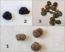
Background: Gallstone illness is a common problem of the biliary system, and its development is known to involve more than one factor. Changes in metabolism, bile composition, and genetic background may all contribute to the formation of gallstones. Objective: The recent work aimed to assess the expression of the bile acid–related genes FXR and CYP7A1 in Iraqi individuals with gallstone illness and to examine their relation to gallstone composition and clinical findings. Methods: A case–control study was carried out on female individuals with gallstone illness and healthy controls. Gene expression of FXR and CYP7A1 was measured using quantitative real-time PCR. Gallstones were examined based on their physical appearance and analyzed via Fourier transform infrared (FTIR) spectroscopy. Clinical and hematological data were collected and statistically analyzed. Results: Individuals showed differences in body mass index and some hematological markers when relative to controls, and a positive family history was more commonly observed. Individuals exhibited reduced expression levels of both FXR and CYP7A1, particularly among individuals with a familial predisposition to gallstones. Gallstone analysis revealed pigment, cholesterol, and mixed types, with mixed stones being the most common form. Conclusions: The results suggested that gallstone illness contributes to alterations in the expression of bile acid-related genes and heterogeneous stone composition. The prevalence of mixed stones seems to indicate that gallstone development follows multiple mechanisms rather than a singular pathway.
Downloads
References
Sun H, Warren J, Yip J, Ji Y, Hao S, Han W, et al. Factors influencing gallstone formation: A review of the literature. Biomolecules. 2022;12(4):550. doi: 10.3390/biom12040550.
Pęczuła A, Czaplicki A, Przybyłkowski A. Genetics of gallstones. Genes. 2025;16(3):256. doi: 10.3390/genes16030256.
Khalaf SK, Al Mousawi J, Hussein A, Al Asadi J. Prevalence and risk factors of asymptomatic gallstones in a sample of population in Basrah, Iraq. Arch Med. 2016;8(4):1-6. doi: 10.21767/1989-5216.1000146.
Al-Obaidi SM, Abdulla TS, Al-Alawi MS, Mohammed HM. The prevalence of silent gallstones and its relation to some risk factors in Iraq. Iraqi Postgrad Med J. 2006;5(2):146-150.
Li H, Zhang C. Association between triglyceride-glucose index and gallstones: a cross-sectional study. Sci Rep. 2024;14(1):17778. doi: s41598-024-68841-6.
Zheng J, Dong H, Wan H, Yang Q, Xu S, Hu T, et al. Positive association between cardiometabolic index and gallstones, with greater impact on women and those younger than 50 years: the NHANES 2017–2020 cross-sectional study. BMC Pub Health. 2025;25(1):2130. doi: 10.1186/s12889-025-23323-w.
Simonsen MH, Erichsen R, Frøslev T, Rungby J, Sørensen HT. Postmenopausal estrogen therapy and risk of gallstone disease: a population-based case–control study. Drug Saf. 2013;36(12):1189-1197. doi: 10.1007/s40264-013-0100-5.
Parra-Landazury N, Cordova-Gallardo J, Méndez-Sánchez N. Obesity and gallstones. Visc Med. 2021;37(5):394–402. doi: 10.1159/000519148.
Lee G, Suh JY, Kim J, Park TY, Do JH, Choi YS, et al. Prevalence of cholesterol gallstones in a Korean population over a 14-year period. Korean J Intern Med. 2025;40(4):584-591. doi: 10.3904/kjim.2025.090.
Singh VK, Jaswal BS, Sharma J, Rai PK. Analysis of stones formed in the human gall bladder and kidney using advanced spectroscopic techniques. Biophys Rev. 2020;12(3):647-668. doi: 10.1007/s12551-020-00672-4.
Ali S, Rasul S, Dawani S, Zahid S, Hussain S, Sarwar O, et al. Effects of chemical composition of cholesterol and pigment stones on the gallbladder mucosa. J Bahria Univ Med Dent Coll. 2022;12(02):68-72.
Dowais R, Al Sharie S, Araydah M, Al Khasawneh S, Haddad F, AlJaiuossi A. Pearl-white gallstones: A report of a case and a chemical analysis via FTIR and XRD. Int J Surg Case Rep. 2021;87:106449. doi: 10.1016/j.ijscr.2021.106449.
Arrout A, El Ghallab Y, Hirri A, Aït Mouss R, Yamari I, Lefriyekh MR, et al. Prediction of cholesterol content in gallstones via FTIR spectroscopy coupled with chemometric tools. Microchem J. 2024;199:109956. doi: 10.1016/j.microc.2024.109956.
Wang Y, Xu H, Zhou X, Chen W, Zhou H. Dysregulated bile acid homeostasis: unveiling its role in metabolic diseases. Med Rev. 2024;4(4):262-283. doi: 10.1515/mr-2024-0020.
Chiang JY, Ferrell JM, Wu Y, Boehme S. Bile acid and cholesterol metabolism in atherosclerotic cardiovascular disease and therapy. Cardiol Plus. 2020;5(4):159-170. doi: 10.4103/2470-7511.305419.
Chiang JY, Ferrell JM. Up to date on cholesterol 7 alpha-hydroxylase (CYP7A1) in bile acid synthesis. Liver Res. 2020;4(2):47-63. doi: 10.1016/j.livres.2020.02.003.
Sikkandar S, Jayakumar S, Gunasekaran S, Renugadevi T, Alwar B. Study on the analysis of human gallstones using Fourier transform infrared spectroscopic technique. Int J ChemTech Res. 2011;3(1):149-154.
Ha BJ, Park S. Classification of gallstones using Fourier-transform infrared spectroscopy and photography. Biomaterials Res. 2018;22(1):18. doi: 10.1186/s40824-018-0139-0.
Bilagi A, Godhi AS. An analytical study of gallstones via Fourier transform infrared spectroscopy technique. Int Surg J. 2022;9(3):584-589. doi: 10.18203/2349-2902.isj20220625.
Raman RG, Selvaraju R. FTIR spectroscopic analysis of human gallstones. Rom J Biophys. 2008;18(4):309-316.
Livak KJ, Schmittgen TD. Analysis of relative gene expression data using real-time quantitative PCR and the 2− ΔΔCT method. Methods. 2001;25(4):402-408. doi: 10.1006/meth.2001.1262.
Portincasa P, Di Ciaula A, Bonfrate L, Stella A, Garruti G, Lamont JT. Metabolic dysfunction-associated gallstone disease: expecting more from critical care manifestations. Intern Emergency Med. 2023;18(7):1897-1918. doi: 10.1007/s11739-023-03185-5.
Fu C, Chen J, Wang Y, Yang Y, Li X, Liu K. Association between complete blood cell count-derived inflammatory biomarkers and gallstones prevalence in American adults under 60 years of age. Front Immunol. 2025;15:1497068. doi: 10.3389/fimmu.2025.1497068.
Parra-Landazury NM, Cordova-Gallardo J, Méndez-Sánchez N. Obesity and gallstones. Visc Med. 2021;37(5):394-402. doi: 10.1159/000519148.
Wang K, Liu Z, Tang R, Sha Y, Wang Z, Chen Y, et al. Gallstones in the era of metabolic syndrome: pathophysiology, risk prediction, and management. Cureus. 2025;17(3). doi: 10.7759/cureus.80541.
Costa CJ, Nguyen MTT, Vaziri H, Wu GY. Genetics of gallstone disease and their clinical significance: A narrative review. J Clin Transl Hepatol. 2024;12(3):316. doi: 10.14218/JCTH.2023.00563.
Fuchs CD, Simbrunner B, Baumgartner M, Campbell C, Reiberger T, Trauner M. Bile acid metabolism and signalling in liver disease. J Hepatol. 2025;82(1):134-153. doi: 10.1016/j.jhep.2024.09.012.
Ye X, Huang D, Dong Z, Wang X, Ning M, Xia J, et al. FXR signaling-mediated bile acid metabolism is critical for alleviation of cholesterol gallstones via lactobacillus strains. Microbiol Spectrum. 2022;10(5):e00518-22. doi: 10.1128/spectrum.00518-22.
Li X, Yin X, Xu J, Geng L, Liu Z. Relationship between abnormal lipid metabolism and gallstone formation. Korean J Gastroenterol. 2025;85(1):11-21. doi: 10.4166/kjg.2024.135.
Shaltout AA, Seoudi R, Almalawi DR, Abdellatief M, Tanthanuch W. Quantitative phase analysis and molecular structure of human gallstones using synchrotron radiation X-ray diffraction and FTIR spectroscopy. Spectrochim Acta Part A Mol Biomol Spectroscopy. 2024;308:123777. doi: 10.1016/j.saa.2024.123777.
Higashizono K, Nakatani E, Hawke P, Fujimoto S, Oba N. Risk factors for gallstone disease onset in Japan: Findings from the Shizuoka Study, a population-based cohort study. Plos One. 2022;17(12):e0274659. doi: 10.1371/journal.pone.0274659.
Ravi PC, Thugu TR, Singh J, Dasireddy RR, Kumar SA, Isaac NV, et al. Gallstone disease and its correlation with thyroid disorders: a narrative review. Cureus. 2023;15(9). doi: 10.7759/cureus.45116.
Downloads
Published
How to Cite
Issue
Section
License
Copyright (c) 2026 Al-Rafidain Journal of Medical Sciences ( ISSN 2789-3219 )

This work is licensed under a Creative Commons Attribution-NonCommercial-ShareAlike 4.0 International License.
Published by Al-Rafidain University College. This is an open access journal issued under the CC BY-NC-SA 4.0 license (https://creativecommons.org/licenses/by-nc-sa/4.0/).


