Giant Serous Cystadenoma of The Pancreatic Tail: Diagnostic Significance of CT Scanning in a Rare Case
DOI:
https://doi.org/10.54133/ajms.v10i2.2794Keywords:
Computed tomography, Mucinous cystadenoma , Neoplastic pancreatic cyst , Pancreatic cyst , Serous cystadenomaAbstract
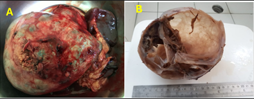
Giant serous cystadenoma (SCA) of the pancreas is a rare benign tumor that typically remains asymptomatic and undetected until it reaches a considerable size, highlighting the diagnostic challenge in differentiating it from other pancreatic cystic lesions. This study seeks to present an uncommon instance of giant pancreatic SCA and underscore the diagnostic and therapeutic methodology. A case report design was used involving a 35-year-old female patient presenting with a painless epigastric mass without associated gastrointestinal or systemic symptoms. Physical examination revealed a firm, non-tender mass in the left upper abdomen. Diagnostic imaging using contrast-enhanced computed tomography (CT) identified a 15 cm multiloculated cystic lesion with thin septations and central calcifications in the body and tail of the pancreas. Surgical intervention consisted of distal pancreatectomy and splenectomy. Histopathological examination confirmed a diagnosis of serous cystadenoma confined to the pancreatic tail, characterized by multiple cystic spaces lined with cuboidal epithelium and clear cytoplasm. The patient experienced an uneventful recovery and was discharged on the fifth postoperative day. This case underscores the critical role of radiologic evaluation in differentiating SCAs from other cystic lesions such as pseudocysts and mucinous cystadenomas to support accurate preoperative planning. The findings suggest that, despite their benign nature, early recognition and surgical management of large SCAs are essential to preventing complications, and improved awareness is needed to enhance diagnostic accuracy and avoid overtreatment or delayed intervention.
Downloads
References
Basturk O, Coban I, Adsay NV. Pancreatic cysts: pathologic classification, differential diagnosis, and clinical implications. Arch Pathol Lab Med. 2009;133(3):423-438. doi: 10.5858/133.3.423. DOI: https://doi.org/10.5858/133.3.423
Chalhoub N, Mourad C, Mouaccadieh L, El Khoury E. Serous cystadenoma of the pancreas: radio-anatomical correlation. Int J Radiol Radiat Ther. 2017;4(1):326-328. doi: 10.15406/ijrrt.2017.04.00087. DOI: https://doi.org/10.15406/ijrrt.2017.04.00087
Chu LC, Singhi AD, Haroun RR, Hruban RH, Fishman EK. The many faces of pancreatic serous cystadenoma: Radiologic and pathologic correlation. Diagn Interv Imaging. 2017;98(3):191–202. doi: 10.1016/j.diii.2016.08.005. DOI: https://doi.org/10.1016/j.diii.2016.08.005
Kim YH, Saini S, Sahani D, Hahn PF, Mueller PR, Auh YH. Imaging diagnosis of cystic pancreatic lesions: pseudocyst versus nonpseudocyst. Radiographics. 2005;25(3):671-685. doi: 10.1148/rg.253045104. DOI: https://doi.org/10.1148/rg.253045104
Tseng JF, Warshaw AL, Sahani DV, Lauwers GY, Rattner DW, Fernandez-del Castillo C. Serous cystadenoma of the pancreas: tumor growth rates and recommendations for treatment. Ann Surg. 2005;242(3):413-419; discussion 419-421. doi: 10.1097/01.sla.0000179651.21193.2c. DOI: https://doi.org/10.1097/01.sla.0000179651.21193.2c
Bramis K, Petrou A, Papalambros A, Manzelli A, Mantonakis E, Brennan N, et al. Serous cystadenocarcinoma of the pancreas: report of a case and management reflections. World J Surg Oncol. 2012;10:51. doi: 10.1186/1477-7819-10-51. DOI: https://doi.org/10.1186/1477-7819-10-51
Downloads
Published
How to Cite
Issue
Section
License
Copyright (c) 2026 Al-Rafidain Journal of Medical Sciences ( ISSN 2789-3219 )

This work is licensed under a Creative Commons Attribution-NonCommercial-ShareAlike 4.0 International License.
Published by Al-Rafidain University College. This is an open access journal issued under the CC BY-NC-SA 4.0 license (https://creativecommons.org/licenses/by-nc-sa/4.0/).


