Anomalous Bilateral DeLano Type III Trans-Sphenoidal Optic Nerve Course in an Iraqi Patient
DOI:
https://doi.org/10.54133/ajms.v10i1.2777Keywords:
Arabs, Congenital anomalies, Functional endoscopic sinus surgery, Paranasal sinuses, Sphenoid bone, Visual pathwaysAbstract
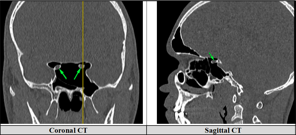
Anomalous trans-sphenoidal course of the optic nerve represents a rare anatomical variant with important clinical implications. Because the optic nerve may bulge into or traverse the sphenoid sinus in the setting of extensive sphenoid sinus pneumatization, such unrecognized anomalies increase the risk of optic nerve or visual pathway injuries during endoscopic or surgical procedures. Early recognition on preoperative imaging is therefore crucial. We are presenting a case study of an eighteen-year-old Iraqi male who underwent a paranasal computed tomography at Baghdad Medical City; clinical history and presenting complaint were unavailable. Imaging demonstrated marked sino-nasal asymmetry with a severe left-sided nasal septum deviation, a larger right inferior turbinate, mild left Schneiderian membrane thickening, an asymmetric multi-septate sphenoid sinus with a left lateral recess, bilateral anterior clinoid process pneumatization, and small right Onodi cells. Most notably, both optic nerves followed an anomalous DeLano type III trans-sphenoidal course, which is an uncommon but clinically significant sino-nasal anatomic variant. Careful preoperative assessment of sphenoid sinus pneumatization, septation, and optic-nerve relationships is essential to prevent iatrogenic optic-nerve injury and to guide endoscopic sinus and skull-base procedures, including functional endoscopic sinus surgery (FESS). Radiology reports should explicitly document these variants, and significant findings should prompt multidisciplinary discussion.
Downloads
References
Baloiu AI, Filipoiu F, Toader C, Covache-Busuioc RA, Munteanu O, Serban M. Sphenoid sinus hyperpneumatization: anatomical variants, molecular blueprints, and AI-augmented roadmaps for skull base surgery. Front Endocrinol (Lausanne). 2025;16:1634206. doi: 10.3389/fendo.2025.1634206. DOI: https://doi.org/10.3389/fendo.2025.1634206
Kar M, Bayar Muluk N, Alqunaee M, Manole F, Cingi C. Functional endoscopic sinus surgery: Key points for safer surgery. Ear Nose Throat J. 2024;103(S3):5S-14S. doi: 10.1177/01455613241287280. DOI: https://doi.org/10.1177/01455613241287280
Papadopoulou AM, Chrysikos D, Samolis A, Tsakotos G, Troupis T. Anatomical variations of the nasal cavities and paranasal sinuses: A systematic review. Cureus. 2021;13(1):e12727. doi: 10.7759/cureus.12727. DOI: https://doi.org/10.7759/cureus.12727
Dessi P, Moulin G, Castro F, Chagnaud C, Cannoni M. Protrusion of the optic nerve into the ethmoid and sphenoid sinus: prospective study of 150 CT studies. Neuroradiology. 1994;36(7):515-516. doi: 10.1007/BF00593511. DOI: https://doi.org/10.1007/BF00593511
DeLano MC, Fun FY, Zinreich SJ. Relationship of the optic nerve to the posterior paranasal sinuses: a CT anatomic study. AJNR Am J Neuroradiol. 1996;17(4):669-675. PMID: 8730186.
Fadda GL, Petrelli A, Urbanelli A, Castelnuovo P, Bignami M, Crosetti E, et al. Risky anatomical variations of sphenoid sinus and surrounding structures in endoscopic sinus surgery. Head Face Med. 2022;18(1):29. doi: 10.1186/s13005-022-00336-z. DOI: https://doi.org/10.1186/s13005-022-00336-z
Erdur ZB. Relationship between the protrusion of the optic nerve and internal carotid artery and sphenoid sinus volume. J Acad Res Med. 2023;13(2):113-117. doi: 10.4274/jarem.galenos.2023.88700. DOI: https://doi.org/10.4274/jarem.galenos.2023.88700
Dalati HA, Jabbr MS, Hammadi IS. Uncommon optic nerve course in the sphenoid sinus. Saudi J Med Med Sci. 2017;5(2):175-176. doi: 10.4103/1658-631X.204867. DOI: https://doi.org/10.4103/1658-631X.204867
Yiğit Ö, Acıoğlu E, Çakır ZA, Şişman AS, Barut AY. Concha bullosa and septal deviation. Eur Arch Otorhinolaryngol. 2010;267(9):1397–401. doi: 10.1007/s00405-010-1228-9. DOI: https://doi.org/10.1007/s00405-010-1228-9
Dieguez FL, Rosa CSD, Braz-Silva PH, Lopes SLPDC, Costa ALF. Three-Dimensional Volumetric Investigation of Onodi Cells: A Multi-Slice Computed Tomography Study. Int Arch Otorhinolaryngol. 2024;28(02):e196–202. doi: 10.1055/s-0043-1773762. DOI: https://doi.org/10.1055/s-0043-1773762
Valenzuela-Fuenzalida JJ, Baez-Flores B, Sepúlveda RÁ, Medina CM, Pérez R, López E, et al. Anatomical variations and abnormalities of the maxillary region and clinical implications: A systematic review and meta-analysis. Medicine. 2023;102(38):e34510. doi: 10.1097/MD.0000000000034510. DOI: https://doi.org/10.1097/MD.0000000000034510
Downloads
Published
How to Cite
Issue
Section
License
Copyright (c) 2026 Al-Rafidain Journal of Medical Sciences ( ISSN 2789-3219 )

This work is licensed under a Creative Commons Attribution-NonCommercial-ShareAlike 4.0 International License.
Published by Al-Rafidain University College. This is an open access journal issued under the CC BY-NC-SA 4.0 license (https://creativecommons.org/licenses/by-nc-sa/4.0/).


