Emerging Oxidative Stress Biomarkers and Mitochondrial Dysfunction in Preeclampsia from Pathogenesis to Precision Medicine: A Narrative Review
Keywords:
Biomarker, Mitochondrial dysfunction, Oxidative stress, Pre-eclampsia, Reactive oxygen speciesAbstract
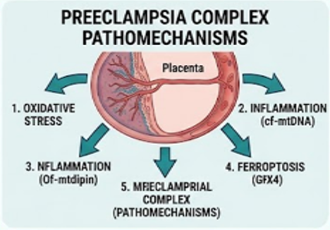
Preeclampsia continues to be a major cause of maternal and neonatal morbidity worldwide. Current diagnostic criteria are based on clinical signs that appear relatively late. Target organ damage is already in progress, and early disease progress often goes undetected at this time. Recent research has altered our perception of this condition and has confirmed that trophoblast mitochondria are actively involved in the etiology of hypertensive disease and not just responding to it. Mitochondrial dysfunction in the placenta results in increased production of free radicals, breakdown of the membrane, disruption in cellular iron balance, and poor organelle clearance, all of which affect the function of the placenta negatively. This review focuses on five important mitochondrial biomarkers that characterize this destructive process: 8-hydroxy-2'-deoxyguanosine indicates direct oxidative damage to mitochondrial DNA, and changes in copy numbers and cell-free DNA fragments in plasma indicate increased cell damage and systemic inflammation. Oxidized cardiolipin and malondialdehyde indicate membrane lipid degradation. Changes in levels of GPX4, mitochondrial ferritin, and transferrin receptor 1 indicate initiation of iron-dependent ferroptosis. Finally, reduced PINK1/Parkin signaling is a sign of the cell's cleanup system failing, allowing damaged mitochondria to accumulate and pushing trophoblasts into premature aging. These markers together depict the convergence of redox imbalance, structural membrane damage, iron dysregulation, and clearance defects driving placental breakdown and maternal disease.
Downloads
References
1. Parrettini S, Caroli A, Torlone E. Nutrition and metabolic adaptations in physiological and complicated pregnancy: Focus on obesity and gestational diabetes. Front Endocrinol (Lausanne). 2020;11. doi: 10.3389/fendo.2020.611929.
2. Chiang YT, Chen JH, Chen KH. The molecular and cellular basis of physiological changes in pregnancy and its implications in neurologic and ophthalmic pathologies. Int J Mol Sci. 2025;26(11):5220. doi: 10.3390/ijms26115220.
3. Chakulya M, Mulambo P, Chama GC, Nalavwe L, Pulukuta IM, Simwaba P, et al. Preeclampsia, prevalence and associated factors. PLoS One. 2025;20(12):e0337190. doi: 10.1371/journal.pone.0337190.
4. Kutllovci Hasani K, Ajeti N, Goswami N. Understanding preeclampsia: Cardiovascular pathophysiology, histopathological insights and molecular biomarkers. Med Sci. 2025;13(3):154. doi: 10.3390/medsci13030154.
5. Vigil-De Gracia P, Vargas C, Sánchez J, Collantes-Cubas J. Preeclampsia: Narrative review for clinical use. Heliyon. 2023;9(3):e14187. doi: 10.1016/j.heliyon.2023.e14187.
6. Witkowski P, Dzieński B, Stefańska K, Tomaszewicz M, Zabielska-Kaczorowska M, Wydra D. Preeclampsia: Contemporary concepts in pathophysiology, risk stratification, prevention and monitoring. J Clin Med. 2026;15(5):1944. doi: 10.3390/jcm15051944.
7. Turbeville HR, Sasser JM. Preeclampsia beyond pregnancy: long-term consequences for mother and child. Am J Physiol-Renal Physiol. 2020;318(6):F1315–1326. doi: 10.1152/ajprenal.00071.2020.
8. Koulouraki S, Paschos V, Pervanidou P, Christopoulos P, Gerede A, Eleftheriades M. Short- and long-term outcomes of preeclampsia in offspring: Review of the literature. Children. 2023;10(5):826. doi: 10.3390/children10050826.
9. Vornic I, Buciu V, Furau CG, Gaje PN, Ceausu RA, Dumitru CS, et al. Oxidative stress and placental pathogenesis: A contemporary overview of potential biomarkers and emerging therapeutics. Int J Mol Sci. 2024;25(22):12195. doi: 10.3390/ijms252212195.
10. Parapob N, Luewan S, Kamlungkuea T, Tongsong T. Oxidative stress in pathogenesis of preeclampsia: Mechanistic and clinical insights. Antioxidants. 2026;15(3):387. doi: 10.3390/antiox15030387.
11. Chiarello DI, Abad C, Rojas D, Toledo F, Vázquez CM, Mate A, et al. Oxidative stress: Normal pregnancy versus preeclampsia. Biochim Biophys Acta. 2020;1866(2):165354. doi: 10.1016/j.bbadis.2018.12.005.
12. Backston K, Morgan J, Patel S, Koka R, Hu J, Raina R. Oxidative stress and endothelial dysfunction: The pathogenesis of pediatric hypertension. Int J Mol Sci. 2025;26(11):5355. doi: 10.3390/ijms26115355.
13. Abu-Awwad SA, Craina M, Boscu L, Bernad E, Ciordas PD, Marian C, et al. Lipid profile variations in pregnancies with and without cardiovascular risk: Consequences for both mother and newborn. Children. 2023;10(9):1521. doi: 10.3390/children10091521.
14. Chen YS, Tian HX, Rong DC, Wang L, Chen S, Zeng J, et al. ROS homeostasis in cell fate, pathophysiology, and therapeutic interventions. Mol Biomed. 2025;6(1):89. doi: 10.1186/s43556-025-00338-8.
15. Meena P, Batool A. Electrolyte homeostasis in pregnancy: from physiological adaptations to clinical disturbances—A nephrologist’s perspective. Front Nephrol. 2026;6. doi: 10.3389/fneph.2026.1773415.
16. Kornacki J, Olejniczak O, Sibiak R, Gutaj P, Wender-Ożegowska E. Pathophysiology of pre-eclampsia—Two theories of the development of the disease. Int J Mol Sci. 2023;25(1):307. doi: 10.3390/ijms25010307.
17. Greenbaum S, Averbukh I, Soon E, Rizzuto G, Baranski A, Greenwald NF, et al. A spatially resolved timeline of the human maternal–fetal interface. Nature. 2023;619(7970):595–605. doi: 10.1038/s41586-023-06298-9.
18. Gusella A, Martignoni G, Giacometti C. Behind the curtain of abnormal placentation in pre-eclampsia: From molecular mechanisms to histological hallmarks. Int J Mol Sci. 2024;25(14):7886. doi: 10.3390/ijms25147886.
19. Rana S, Burke SD, Karumanchi SA. Imbalances in circulating angiogenic factors in the pathophysiology of preeclampsia and related disorders. Am J Obstet Gynecol. 2022;226(2):S1019–1034. doi: 10.1016/j.ajog.2020.10.022.
20. Smith AN, Wang X, Thomas DG, Tatum RE, Booz GW, Cunningham MW. The role of mitochondrial dysfunction in preeclampsia: Causative factor or collateral damage? Am J Hypertens. 2021;34(5):442–452. doi: 10.1093/ajh/hpab003.
21. Hu XQ, Zhang L. Mitochondrial dysfunction in the pathogenesis of preeclampsia. Curr Hypertens Rep. 2022;24(6):157–172. doi: 10.1007/s11906-022-01184-7.
22. Zhou X, Zhao X, Zhou W, Qi H, Zhang H, Han TL, et al. Impaired placental mitophagy and oxidative stress are associated with dysregulated BNIP3 in preeclampsia. Sci Rep. 2021;11(1):20469. doi: 10.1038/s41598-021-99837-1.
23. Green DR. The mitochondrial pathway of apoptosis. Cold Spring Harb Perspect Biol. 2022;14(5):a041038. doi: 10.1101/cshperspect.a041038.
24. Vasilaki I, Potiris A, Moustakli E, Mavrogianni D, Daponte N, Karampitsakos T, et al. Linking oxidative stress to placental dysfunction: The key role of mitochondria in trophoblast function. Med Sci. 2026;14(1):53. doi: 10.3390/medsci14010053
25. Kajdy A, Modzelewski J, Cymbaluk-Płoska A, Kwiatkowska E, Bednarek-Jędrzejek M, Borowski D, et al. Molecular pathways of cellular senescence and placental aging in late fetal growth restriction and stillbirth. Int J Mol Sci. 2021;22(8):4186. doi: 10.3390/ijms22084186.
26. Čepelak I, Dodig S, Dodig DČ. Ferroptosis: regulated cell death. Arch Indust Hyg Toxicol. 2020;71(2):99–109. doi: 10.2478/aiht-2020-71-3366.
27. Sangkhae V, Fisher AL, Ganz T, Nemeth E. Iron homeostasis during pregnancy: Maternal, placental, and fetal regulatory mechanisms. Annu Rev Nutr. 2023;43(1):279–300. doi: 10.1146/annurev-nutr-061021-030404.
28. Zheng Y, Sun J, Luo Z, Li Y, Huang Y. Emerging mechanisms of lipid peroxidation in regulated cell death and its physiological implications. Cell Death Dis. 2024;15(11):859. doi: 10.1038/s41419-024-07244-x.
29. Barbouti A, Varvarousis DN, Kanavaros P. The role of oxidative stress-induced senescence in the pathogenesis of preeclampsia. Antioxidants. 2025;14(5):529. doi: 10.3390/antiox14050529.
30. Sultana Z, Maiti K, Dedman L, Smith R. Is there a role for placental senescence in the genesis of obstetric complications and fetal growth restriction? Am J Obstet Gynecol. 2018;218(2):S762–773. doi: 10.1016/j.ajog.2017.11.567.
31. Wang C, Zhang M. Neutrophil extracellular traps in pregnancy complications. Front Immunol. 2026;17. doi: 10.3389/fimmu.2026.1798749.
32. Hernández González LL, Pérez-Campos Mayoral L, Hernández-Huerta MT, Mayoral Andrade G, Martínez Cruz M, Ramos-Martínez E, et al. Targeting neutrophil extracellular trap formation: Exploring promising pharmacological strategies for the treatment of preeclampsia. Pharmaceuticals. 2024;17(5):605. doi: 10.3390/ph17050605.
33. Chen WA, Boskovic DS. Neutrophil extracellular DNA traps in response to infection or inflammation, and the roles of platelet interactions. Int J Mol Sci. 2024;25(5):3025. doi: 10.3390/ijms25053025.
34. Hu XQ, Zhang L. Hypoxia and mitochondrial dysfunction in pregnancy complications. Antioxidants. 2021;10(3):405. doi: 10.3390/antiox10030405.
35. Hu XQ, Zhang L. Hypoxia and the integrated stress response promote pulmonary hypertension and preeclampsia: Implications in drug development. Drug Discov Today. 2021;26(11):2754–2773. doi: 10.1016/j.drudis.2021.07.011.
36. Al-Ogaidi SO, Abdulsattar SA, Al-Dulaimi HM. FTO rs17817449 gene polymorphism as a predictor for maternal obesity in Iraqi pregnant women. Indian J Public Health Res Dev. 2019;10(4):678. doi: 10.5958/0976-5506.2019.00780.0.
37. Zhang H, Burr SP, Chinnery PF. The mitochondrial DNA genetic bottleneck: inheritance and beyond. Essays Biochem. 2018;62(3):225–234. doi: 10.1042/EBC20170096.
38. Gustafsson CM, Falkenberg M, Larsson NG. Maintenance and expression of mammalian mitochondrial DNA. Annu Rev Biochem. 2016;85(1):133–160. doi: 10.1146/annurev-biochem-060815-014402.
39. Liu L, Wang A, Yi K. Causal relationships between mitochondrial DNA copy number and hypertensive disorders in pregnancy: A Mendelian randomization study. Eurasian J Med Oncol. 2025;10(1):100. doi: 10.36922/EJMO025140077.
40. Abdulsattar SA, Nori W, Mohammed EA. Biomarker-guided prediction of pregnancy viability, current evidence and emerging insights: a narrative review. Al-Rafidain J Med Sci. 2025;9(1):246-254. doi: 10.54133/ajms.v9i1.2300.
41. Pandey D, Yevale A, Naha R, Kuthethur R, Chakrabarty S, Satyamoorthy K. Mitochondrial DNA copy number variation – A potential biomarker for early onset preeclampsia. Pregnancy Hypertens. 2021;23:1–4. doi: 10.1016/j.preghy.2020.10.002.
42. Toledano JM, Puche-Juarez M, Galvez-Navas JM, Moreno-Fernandez J, Diaz-Castro J, Ochoa JJ. Pregnancy disorders: A potential role for mitochondrial altered homeostasis. Antioxidants. 2024;13(8):979. doi: 10.3390/antiox13080979.
43. Kamar Bashah NA, Hamid AA, Adam SH, Jaffar FHF, Abd Rahman IZ, Mokhtar MH. Role of AMP-activated protein kinase (AMPK) in female reproduction: A review. Int J Mol Sci. 2025;26(14):6833. doi: 10.3390/ijms26146833.
44. Abu Shelbayeh O, Arroum T, Morris S, Busch KB. PGC-1α is a master regulator of mitochondrial lifecycle and ROS stress response. Antioxidants. 2023;12(5):1075. doi: 10.3390/antiox12051075.
45. Cushen SC, Ricci CA, Bradshaw JL, Silzer T, Blessing A, Sun J, et al. Reduced maternal circulating cell‐free mitochondrial DNA is associated with the development of preeclampsia. J Am Heart Assoc. 2022;11(2). doi: 10.1161/JAHA.121.021726.
46. Vasilaki I, Potiris A, Moustakli E, Mavrogianni D, Daponte N, Karampitsakos T, et al. Linking oxidative stress to placental dysfunction: The key role of mitochondria in trophoblast function. Med Sci. 2026;14(1):53. doi: 10.3390/medsci14010053.
47. Paradies G, Paradies V, Ruggiero FM, Petrosillo G. Role of cardiolipin in mitochondrial function and dynamics in health and disease: Molecular and pharmacological sspects. Cells. 2019;8(7):728. doi: 10.3390/cells8070728.
48. Venkatraman K, Budin I. Cardiolipin remodeling maintains the inner mitochondrial membrane in cells with saturated lipidomes. J Lipid Res. 2024;65(8):100601. doi: 10.1016/j.jlr.2024.100601.
49. Petrosillo G, Ruggiero FM, Paradies G. Role of reactive oxygen species and cardiolipin in the release of cytochrome c from mitochondria. FASEB J. 2003;17(15):2202–2208. doi: 10.1096/fj.03-0012com.
50. Goswami D, Tannetta DS, Magee LA, Fuchisawa A, Redman CWG, Sargent IL, et al. Excess syncytiotrophoblast microparticle shedding is a feature of early-onset pre-eclampsia, but not normotensive intrauterine growth restriction. Placenta. 2006;27(1):56–61. doi: 10.1016/j.placenta.2004.11.007.
51. Afrose D, Chen H, Ranashinghe A, Liu C, Henessy A, Hansbro PM, et al. The diagnostic potential of oxidative stress biomarkers for preeclampsia: systematic review and meta-analysis. Biol Sex Differ. 2022;13(1):26. doi: 10.1186/s13293-022-00436-0.
52. Khoubnasabjafari M, Ansarin K, Jouyban A. Variations of malondialdehyde in pre-eclampsia. Hypertens Pregnancy. 2016;35(3):346–349. doi: 10.3109/10641955.2016.1148163.
53. Chen Z, Gan J, Zhang M, Du Y, Zhao H. Ferroptosis and its emerging role in pre-eclampsia. Antioxidants. 2022;11(7):1282. doi: 10.3390/antiox11071282.
54. Chen Y, Fang ZM, Yi X, Wei X, Jiang DS. The interaction between ferroptosis and inflammatory signaling pathways. Cell Death Dis. 2023;14(3):205. doi: 10.1038/s41419-023-05716-0.
55. Peng X, Lin Y, Li J, Liu M, Wang J, Li X, et al. Evaluation of glutathione peroxidase 4 role in preeclampsia. Sci Rep. 2016;6(1):33300. doi: 10.1038/srep33300.
56. Bucarey JL, Casas M, Espinosa A. Mitochondrial iron handling and lipid peroxidation as drivers of ferroptosis. Int J Mol Sci. 2026;27(5):2232. doi: 10.3390/ijms27052232.
57. Jin J, Xu Z, Liu Z, Huang L, Liu F, Liu M, et al. Association between maternal iron status and the risk of pre-eclampsia: a case-control study. Asia Pac J Clin Nutr. 2024;33(2):184–193. doi: 10.6133/apjcn.202406_33(2).0005.
58. Yang Y, Zuo H, Ma X, Chen W, Sun M, Xiang Q, et al. Iron overload-induced ferroptosis drives placental dysfunction in preeclampsia. Hypertension. 2026;83(7). doi: 10.1161/HYPERTENSIONAHA.125.26344.
59. Zhong J, Jiang R, Liu N, Cai Q, Cao Q, Du Y, et al. Iron–immune crosstalk at the maternal–fetal interface: Emerging mechanisms in the pathogenesis of preeclampsia. Antioxidants. 2025;14(7):890. doi: 10.3390/antiox14070890.
60. Yildirim RM, Ergun Y, Basar M. Mitochondrial dysfunction, mitophagy and their correlation with perinatal complications: Preeclampsia and low birth weight. Biomedicines. 2022;10(10):2539. doi: 10.3390/biomedicines10102539.
61. Zhang Z, Zhang M, Jin H, Lv S, Li Y, Li Y. Mitochondrial quality control and cell death. Int J Mol Sci. 2025;26(22):11084. doi: 10.3390/ijms262211084.
62. Sauvé V, Stefan E, Croteau N, Goiran T, Fakih R, Bansal N, et al. Activation of parkin by a molecular glue. Nat Commun. 2024;15(1):7707. doi: 10.1038/s41467-024-51889-3.
63. Sun Y, Lv D, Xie Y, Xu H, Li X, Li F, et al. PINK1-mediated mitophagy induction protects against preeclampsia by decreasing ROS and trophoblast pyroptosis. Placenta. 2023;143:1–11. doi: 10.1016/j.placenta.2023.09.010.
64. Vaka R, Deer E, LaMarca B. Is mitochondrial oxidative stress a viable therapeutic target in preeclampsia? Antioxidants. 2022;11(2):210. doi: 10.3390/antiox11020210.
65. Voros C, Stavros S, Sapantzoglou I, Mavrogianni D, Daskalaki MA, Theodora M, et al. The role of placental mitochondrial dysfunction in adverse perinatal outcomes: A systematic review. J Clin Med. 2025;14(11):3838. doi: 10.3390/jcm14113838.
66. Nori W, Helmi ZR. Can follicular fluid 8-oxo-2′-deoxyguanosine predict the clinical outcomes in ICSI cycle among couples with normospermia male? Obstet Gynecol Sci. 2023;66(5):430-440. doi: 10.5468/ogs.22170.
67. Brunst KJ, Zhang L, Zhang X, Baccarelli AA, Bloomquist T, Wright RJ. Associations between maternal lifetime stress and placental mitochondrial DNA mutations in an urban multiethnic cohort. Biol Psychiatry. 2021;89(6):570–578. doi: 10.1016/j.biopsych.2020.09.013.
68. Cushen SC, Sprouse ML, Blessing A, Sun J, Jarvis SS, Okada Y, et al. Cell-free mitochondrial DNA increases in maternal circulation during healthy pregnancy: a prospective, longitudinal study. Am J Physiol-Regul Integrative Comparative Physiol. 2020;318(2):R445–452. doi: 10.1152/ajpregu.00324.2019.
69. Afrose D, Alfonso-Sánchez S, McClements L. Targeting oxidative stress in preeclampsia. Hypertens Pregnancy. 2025;44(1). doi: 10.1080/10641955.2024.2445556.
70. Anto EO, Roberts P, Turpin CA, Wang W. Oxidative stress as a key signaling pathway in placental angiogenesis changes in preeclampsia: Updates in pathogenesis, novel biomarkers and therapeutics. Curr Pharmacogenomics Person Med. 2019;16(3):167–181. doi: 10.2174/1875692117666181207120011.
71. Ibrahim A, Khoo MI, Ismail EHE, Hussain NHN, Zin AAM, Noordin L, et al. Oxidative stress biomarkers in pregnancy: a systematic review. Reprod Biol Endocrinol. 2024;22(1):93. doi: 10.1186/s12958-024-01259-x.
72. Zhou X, Zhang H, Wei L, Wang Z, Zhu S, Yu J, et al. Oxidative stress, mitochondrial dysfunction, and ferroptosis in the placenta of gestational diabetes mellitus. J Mol Med. 2026;104(1):8. doi: 10.1007/s00109-025-02629-7.
73. Fukushima K, Murata M, Tsukimori K, Eisuke K, Wake N. 8-Hydroxy-2-deoxyguanosine staining in placenta is associated with maternal serum uric acid levels and gestational age at diagnosis in pre-eclampsia. Am J Hypertens. 2011;24(7):829–834. doi: 10.1038/ajh.2011.40.
74. Takagi Y, Nikaido T, Toki T, Kita N, Kanai M, Ashida T, et al. Levels of oxidative stress and redox-related molecules in the placenta in preeclampsia and fetal growth restriction. Virchows Arch. 2004;444(1):49–55. doi: 10.1007/s00428-003-0903-2.
75. Anto EO, Coall DA, Asiamah EA, Afriyie OO, Addai-Mensah O, Wiafe YA, et al. Placental lesions and differential expression of pro-and anti-angiogenic growth mediators and oxidative DNA damage marker in placentae of Ghanaian suboptimal and optimal health status pregnant women who later developed preeclampsia. PLoS One. 2022;17(3):e0265717. doi: 10.1371/journal.pone.0265717.
76. Qiu C, Hevner K, Abetew D, Enquobahrie DA, Williams MA. Oxidative DNA damage in early pregnancy and risk of gestational diabetes mellitus: A pilot study. Clin Biochem. 2011;44(10–11):804–808. doi: 10.1016/j.clinbiochem.2011.04.023.
77. Palei AC. Cell-free DNA as a potential biomarker for preeclampsia. Expert Rev Mol Diagn. 2021;21(12):1253–1256. doi: 10.1080/14737159.2021.1997593.
78. Amaral LM, Sandrim VC, Kutcher ME, Spradley FT, Cavalli RC, Tanus-Santos JE, et al. Circulating total cell-free DNA levels are increased in hypertensive disorders of pregnancy and associated with prohypertensive factors and adverse clinical outcomes. Int J Mol Sci. 2021;22(2):564. doi: 10.3390/ijms22020564.
79. Rafaeli-Yehudai T, Imterat M, Douvdevani A, Tirosh D, Benshalom-Tirosh N, Mastrolia SA, et al. Maternal total cell-free DNA in preeclampsia and fetal growth restriction: Evidence of differences in maternal response to abnormal implantation. PLoS One. 2018;13(7):e0200360. doi: 10.1371/journal.pone.0200360.
80. Paradies G. Oxidative stress, cardiolipin and mitochondrial dysfunction in nonalcoholic fatty liver disease. World J Gastroenterol. 2014;20(39):14205. doi: 10.3748/wjg.v20.i39.14205.
81. Fiorucci L, Erba F, Santucci R, Sinibaldi F. Cytochrome c interaction with cardiolipin plays a key role in cell apoptosis: Implications for human diseases. Symmetry (Basel). 2022;14(4):767. doi: 10.3390/sym14040767
82. Baş A, Dur R. The relationship between trace element imbalance, oxidative stress, and endothelial dysfunction in preeclampsia diagnosis. BMC Pregnancy Childbirth. 2026;26(1):766. doi: 10.1186/s12884-026-09277-w.
83. Beharier O, Kajiwara K, Sadovsky Y. Ferroptosis, trophoblast lipotoxic damage, and adverse pregnancy outcome. Placenta. 2021;108:32–38. doi: 10.1016/j.placenta.2021.03.007.
84. Liu M, Wu K, Wu Y. The emerging role of ferroptosis in female reproductive disorders. Biomed Pharmacother. 2023;166:115415. doi: 10.1016/j.biopha.2023.115415.
85. Abbasi U, Abbina S, Gill A, Takuechi LE, Kizhakkedathu JN. Role of iron in the molecular pathogenesis of diseases and therapeutic opportunities. ACS Chem Biol. 2021;16(6):945–972. doi: 10.1021/acschembio.1c00122.
86. Salimi Z, Afsharinasab M, Rostami M, Eshaghi Milasi Y, Mousavi Ezmareh SF, Sakhaei F, et al. Iron chelators: as therapeutic agents in diseases. Ann Med Surg. 2024;86(5):2759–2776. doi: 10.1097/MS9.0000000000001717.
87. Ge P, Dawson VL, Dawson TM. PINK1 and Parkin mitochondrial quality control: a source of regional vulnerability in Parkinson’s disease. Mol Neurodegener. 2020;15(1):20. doi: 10.1186/s13024-020-00367-7.
88. Narendra DP, Jin SM, Tanaka A, Suen DF, Gautier CA, Shen J, et al. PINK1 is selectively stabilized on impaired mitochondria to activate parkin. PLoS Biol. 2010;8(1):e1000298. doi: 10.1371/journal.pbio.1000298
89. Sun Y, Lv D, Xie Y, Xu H, Li X, Li F, et al. PINK1-mediated mitophagy induction protects against preeclampsia by decreasing ROS and trophoblast pyroptosis. Placenta. 2023;143:1–11. doi: 10.1016/j.placenta.2023.09.010.
90. Papapanagiotou A, Daskalaki MA, Gargalionis AN, Margoni A, Papapanagiotou A, Daskalaki MA, et al. The role of angiogenetic factors in preeclampsia. Int J Mol Sci. 2025;26(21):10431. doi: 10.3390/ijms262110431.
Downloads
Published
How to Cite
Issue
Section
License
Copyright (c) 2026 Al-Rafidain Journal of Medical Sciences ( ISSN 2789-3219 )

This work is licensed under a Creative Commons Attribution-NonCommercial-ShareAlike 4.0 International License.
Published by Al-Rafidain University College. This is an open access journal issued under the CC BY-NC-SA 4.0 license (https://creativecommons.org/licenses/by-nc-sa/4.0/).